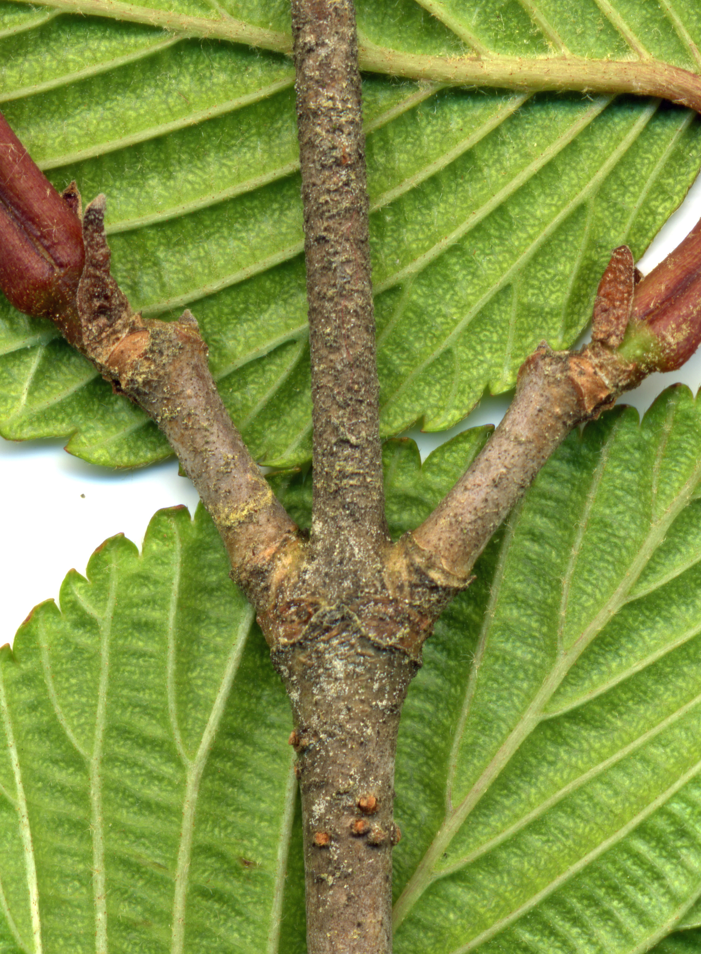
1
To the left is a segment of a branch taken about 18" from the branch's tip.
It is loaded with spores.
In several areas it is so thick that it forms solid masses, making this bush a prolific infection agent!
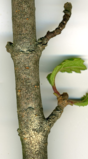
2
Another branch from this bush, again showing the spore density. As before, you can
see that the spore density is by far highest near the branch junctions.
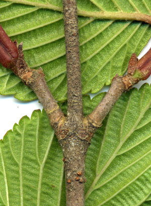
3
The spore density isn't as high here as in the other pictures. Nevertheless, it
again demonstrates the clustering of the spores around the branch junction.
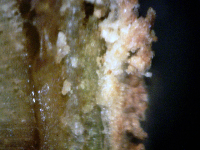
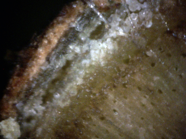
5
Here we can see that the sapwood is infected, and has almost separated from the
heartwood. Furthermore, the bark has become almost totally replaced by white
canker material. (400x)
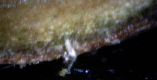
6
The particles of white canker under the bark here are so numerous that they are
pushing the bark out. Careful examination seems to show that each particle
closely resembles a spore in shape. (400x)
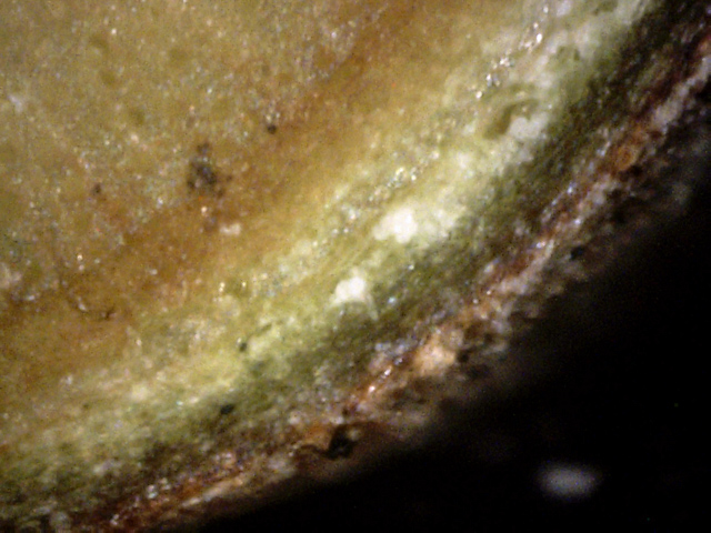
7
This is a rare picture, since it captures white canker material growing from the
phloem, through the dark green cortex, through the outer bark,
transforming into a hypha, and then ending as a young spore.
It ties together all of the infection structures. (400x)
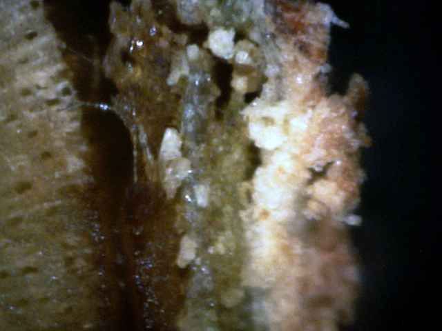
8
The lower-left portion of this photo shows a generally canker-free area under
the bark. However, as you move to the upper-right, canker density rapidly
increases, pushing out the bark slightly. If you look closely, you'll see that
the canker density on the outer bark follows the canker density under the bark. (400x)
9
Finally, this portion of the bark is solidly infected with white canker, so much
so that air spaces have opened up under the bark (400x)
10
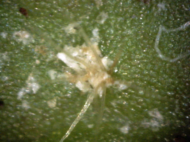
Even infected viburnum leaf tops appear clean. I had to search a bit to find
this area with white canker particles. Note the "octopus" structure. They seem
to be associated with the white canker, but their shape is unlike objects seen
in other infected shrubs or trees. (400x)
11
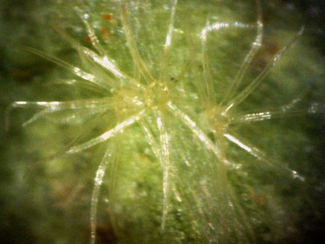
Unlike the relatively clean leaf tops, the leaf bottoms have plenty of these
octopus objects. Here are three of them close together. They each have about 8
"arms". (400x)

12
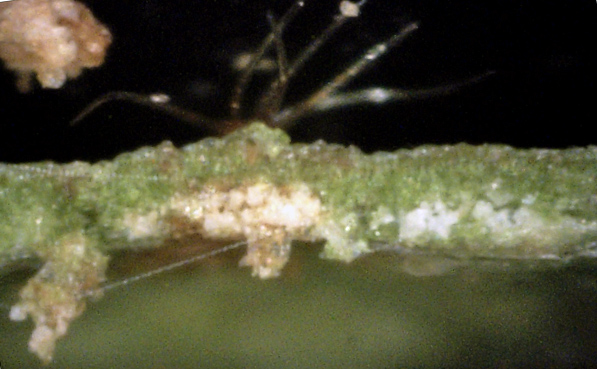
This leaf cross-section explains why viburnum leaves never appear to be infected - the
infection generally resides deep inside the leaf, so can't be seen. The
infection does, however, cause a slight bulge in the leaf. (400x)
13
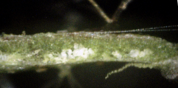
This cross-section shows that the infection became so dense that it pushed out
through the bottom of the leaf. Not only that, but it appears to have caused an
octopus object to arise from the top of the leaf. The brown discoloration may be
because this canker cluser is dying. (400x)
14
Another white canker particle cluster. While the focus is poor, you can make out
an octopus protrusion on the leaf bottom directly below this canker cluster. (400x)
15
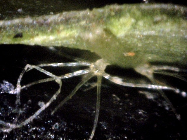
A good view of an octopus object that seems to be feeding off a canker buried
deep within the leaf. This is also a good view of the base of these objects.
This base is a more translucent gray, similar to some canker material. (400x)
This viburnum bush is about 8' tall. The branch shown in these pictures was
taken near the top, at the 6' level. This is the only bush or tree whose leaves
appear to not be affected by this disease. However, some of the stem bark looks
unhealthy, and several years ago there was a bleeding canker at the base of this
bush.
In summary, while white canker can severely affect a viburnum bush, the leaves will
not appear diseased since the white canker material hides deep within the leaf.
However, as with all infected trees and shrubs, the disease easily shows itself
when examining cross-sections of the twigs and leaves using a 400 power microscope.
