
1
The quarter-inch twig shown here was from the tip of a sick-looking rhododendron bush.
Look closely and you will see that this branch is simply loaded with spores!
In fact, it's one of the most prolific spore producers I've seen so far.
The spores seem to prefer year-old wood, and seem to take up residence everywhere along the branch.
The spore density on older wood decreases, probably because the bark there
is virtually falling off due to disease and no more nutrients are available for growth.
2
←
←
↑
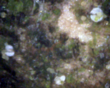
A 400x view showing three spores which appear to be in the act of reproducing.
The arrows show the yellow young spores coming out of the side of the white "mouth".
3
←
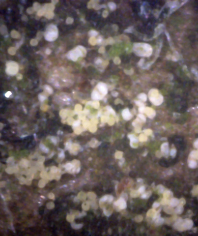
This 400x view of the bark shows an area of abundant spores - young and mature.
The blue arrow points to a white "spider" object in the upper right.
4
↑
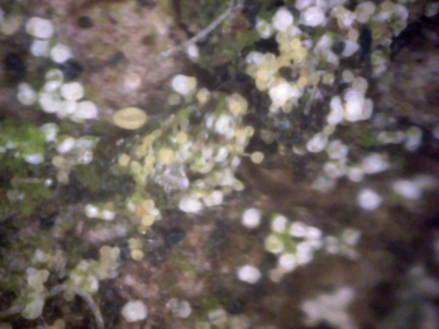
A variety of spore-associated objects is shown in this 400x view of the bark.
Strangely, this view seems to show that the yellow young spores first emerge as green in color
and then evolve into a yellow color.
Also, the blue arrow points to a good view of the often difficult-to-see spore stalk on a newly-born spore.
5
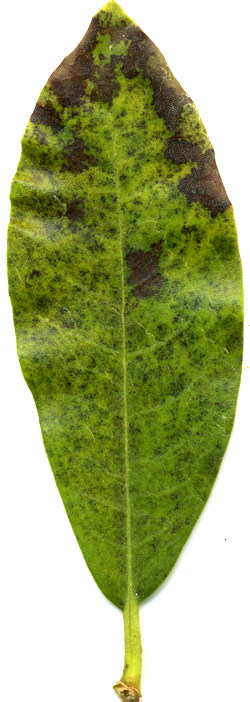
This is a diseased leaf taken from a 10 '' high rhododendron bush on the north side of our garage.
Most other leaves on the bush weren't this bad looking, although they all were somewhat puckered,
as if under attack by some pathogen. Pictures 1 through 9 were all taken
in mid-September 2008.
6
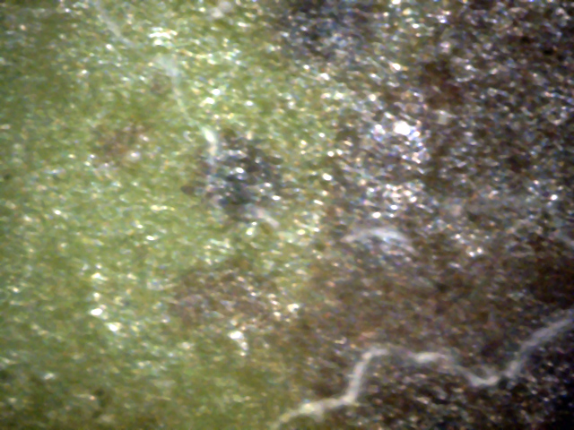
The top side of the rhododendron leaf shown in picture R1-5.
The black areas are dead parts of the leaf.
Note the hyphae near the leaf surface.
They are often associated with these dead spots.
7
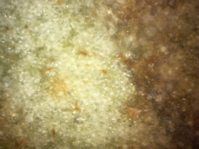
The bottom side of the rhododendron leaf shown in picture 5.
The dark brown area is a dead part of the leaf.
There are what appear to be numerous dead/brown hyphae scattered around.

8
9
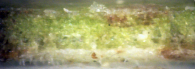
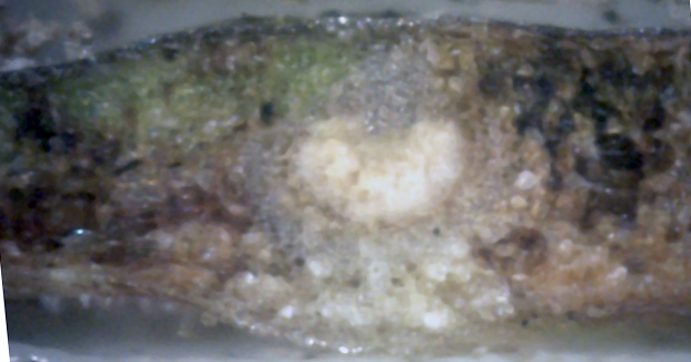
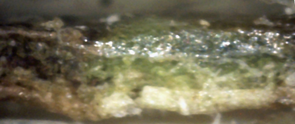
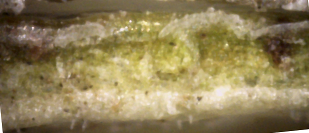
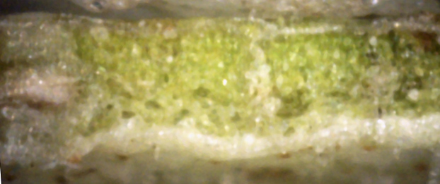
I clamped the leaf shown in picture 5 in a vice, cut it flush with a razor,
and used a 400x microscope to view the interior. Picture set 9 shows 6 of
these edge views. Note that white canker material tends to grow just below the
leaf surface, and predominantly on the bottom surface of the leaf. Look
carefully, and you can see some canker hyphae sticking out of the leaf bottom.
8
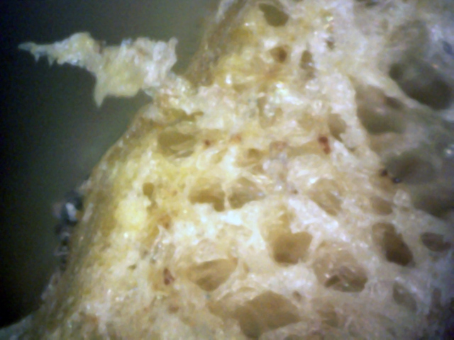
Here is a cross-cut of a 1/4 inch twig from that rhododendron bush.
The cankerous material has consumed a large part of the inner bark. Note the
chunk of it sticking out.
This bush is on the north side of our garage and is now (early June, 2008) full of beautiful red blooms.
The leaves had looked healthy, but during the past week had begun to droop and look sick.