1
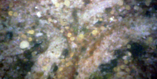
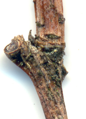
This is a close-up of a branch junction. The spores are in close proximity to it,
although the spore density doesn't appear to be very high.
2
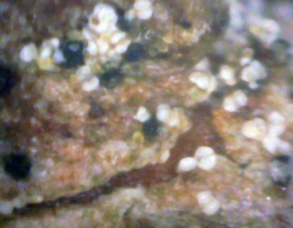
Here is a 400x close-up of the spores at that junction.
Many of these spores have that double-lobed appearance.
Also, many of the spores have a yellow substance between or adjacent to the lobes.

4
Here we see a both spore types - white and yellow.
The two spores in the lower right are interesting because they appear joined and are the same size.
It makes you wonder if the yellow spores are generating the white spores, or vice versa. (400x)
5
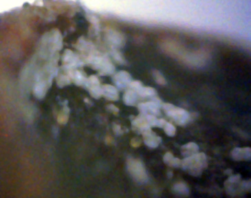
This 400x picture was included simply because it shows the white spores growing on a bark protrusion.
Unfortunately, the microscope's lack of a good depth of view prevented most of it from being in focus.
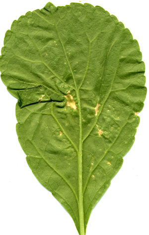
6
Here is what a diseased leaf looks like, with typical white canker puckering (here
squashed flat by the flatbed scanner). The dead areas are usually at the edge of
the leaf, not at the leaf center as here.
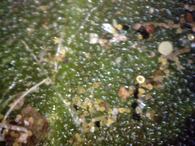
7
This leaftop picture gave me a surprise - I hadn't expected to see young/yellow spores
on the top of any leaf.
Yet, here they were (no spores were on the bottom).
Not only were they there, but almost the entire gamut of spore associated objects were also present!
This picture was taken in early June 2008. When I examined these leaf tops in
mid-September, there wasn't a single yellow spore present, and only a very few
white spores were present. Therefore, the spores were shed in the intervening
months. (400x)
 ←
←
←
←
Pith
←
←
←
←
Pith
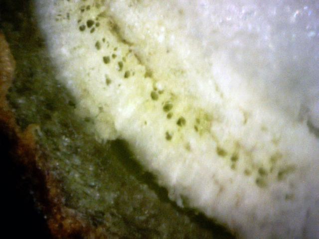
8
Check out the white canker under the inner bark (blue arrow). It matures into
light-gray blobs (red arrows). The black area is a void. The twig's pith is in
the upper-right corner and is a frothy white. (400x)
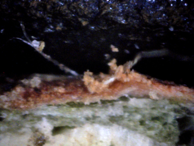
9
Not only is the inner and outer bark riddled with white canker, but two hypha
are also sticking out of the bark. Note particularly the one on the left - it
has given birth to a white canker particle and a young yellow canker spore. This
combination is rarely seen. (400x)
10
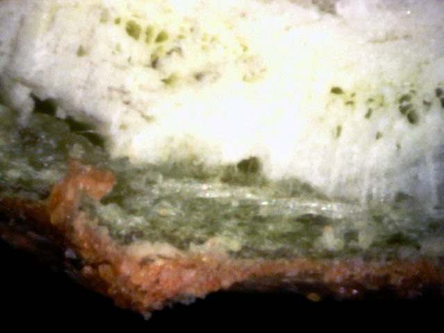
The sapwood and inner bark have white canker growth, and several particles of
white canker can be seen directly under the bulge in the bark. (400x)
11
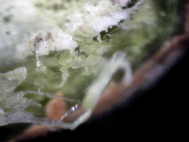
The white canker has grown quite a bit in the sapwood and inner bark, and is now
shooting out hyphae, which look like long icycle-like growths. These growths
seek out other areas to infect. (400x)
 ↓
↓
12
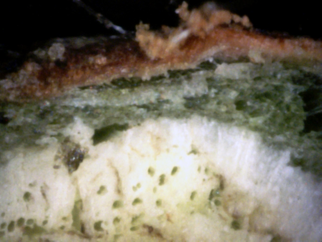
Snow white canker is consuming the sapwood, the inner bark is mostly all canker,
and you can see some white canker particles emerging from the outside of the
bark (red arrow). (400x)
13
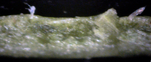
This is a leaf cross-section. Unlike twig cross-sections, it's more difficult to
see obvious signs of white canker in leaf cross-sections. Here we see a piece of
white canker growing up out of the leaf surface, and to the right a hypha
growing out of the surface. (400x)
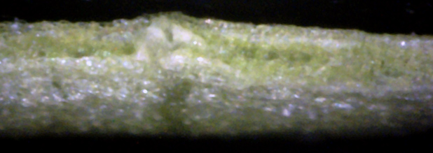
14
The bottom of this leaf seems to have a lot of diffuse white canker material,
and one piece of it is sticking up and almost piercing the top leaf surface,
making a slight bulge in it. (400x)
This vine grows on the south side of our house.
It has generally done well, but white canker took a heavy toll on it the past few years.
Some conclusions we can draw from these pictures:
- White canker yellow spores only appear in late spring/early summer
- White canker appears snow white when growing in the sapwood and chokes off the vessels (pores) there
- White canker is light gray in color when growing within the inner and outer bark
- There are almost no mature white canker spores present in late summer (mid September)
- White canker is easier to detect in twig cross-sections than in leaf cross-sections
