This neighborhood tree has an interesting story associated with it.
I was driving by a home and noticed a tree with only half its normal foliage.
Since it was a Sycamore, and I hadn't checked these trees, I stopped.
The homeowner happened to be in the yard. I'd known him from years ago.
I explained what I was doing and he said I could take whatever branch sample I wanted.
A few minutes later, a tree service truck drove up. Turns out the homeowner was waiting for him.
The homeowner explained that several trees were sick, dropping branches, and wanted them cut down.
The tree service guy happened to look at the sycamore, suggested it was in very bad shape,
and should be cut down! That sure validated my belief that these trees were sick.
2a
2b

2c
2d
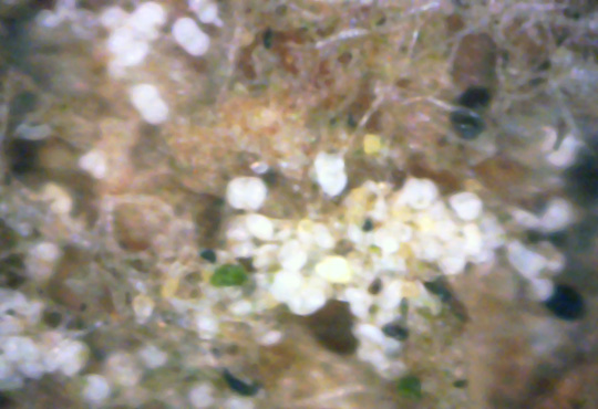
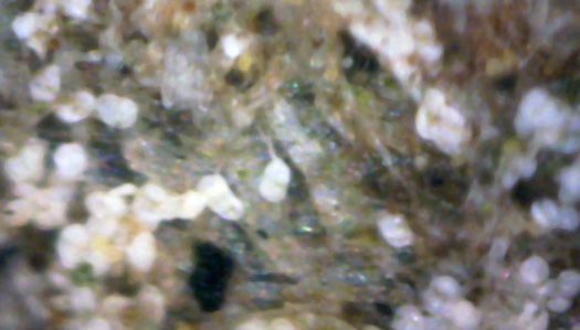
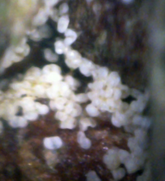
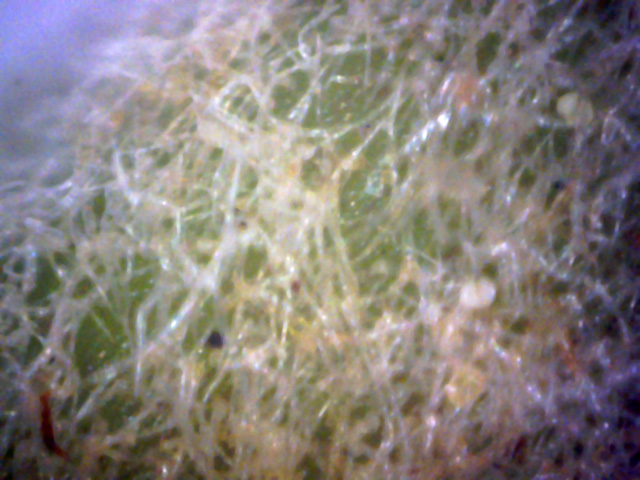
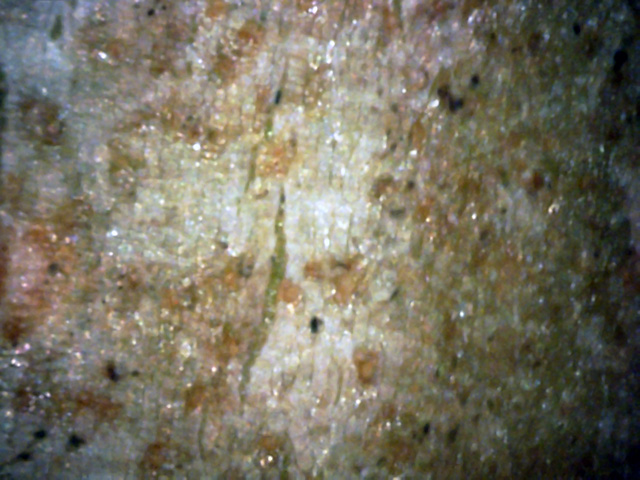
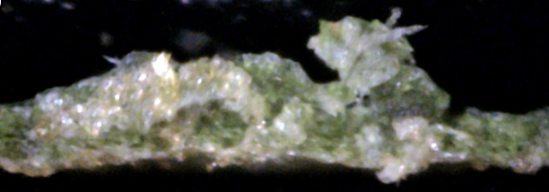
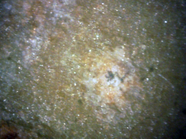
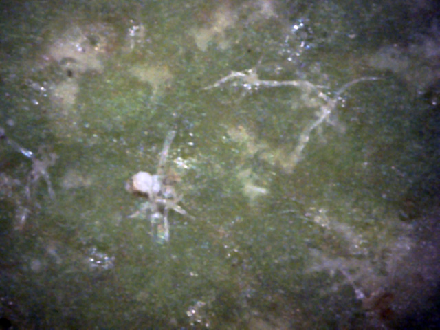
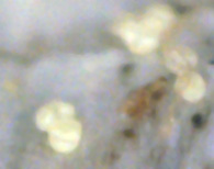
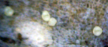
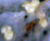
These views show the spores that were on that branch.
Since this was new wood, I wanted to see what very newly implanted spores looked like.
Generally, they were yellowish in color, as in the top picture of the set.
The computer's color correction software tried to make them white in the remaining pictures.
Their yellow color seems to indicate the yellow spores settled in, and then later converted to white spores.
(400x)
3
Unlike other trees, this sycamore tree doesn't seem to have its spores
gather around branch or leaf junctions.
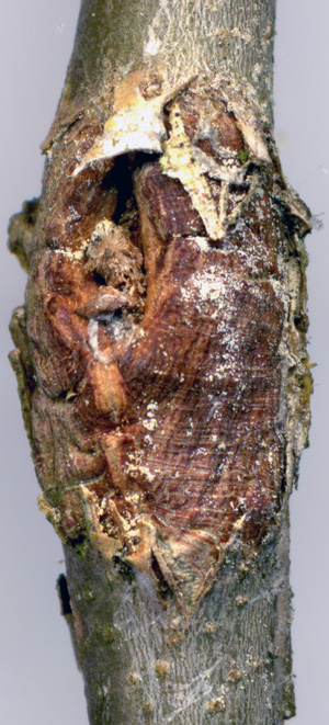
Instead, the spores congregate on wounds, as shown here.
5
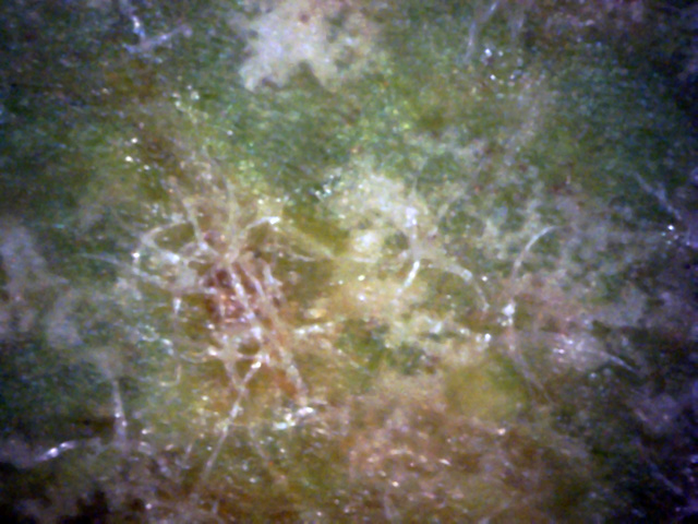
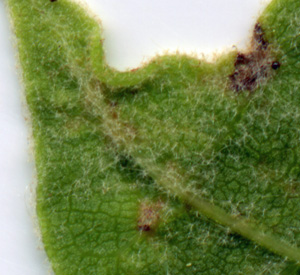
But the big surprise was a sick looking terminal leaf, a portion of which is shown here.
It had a dull look. In fact, it looked hairy. The sickest parts of the leaf had the most hair. (400x)
6a
6b
6c
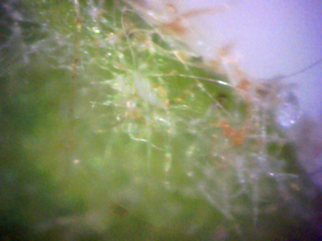
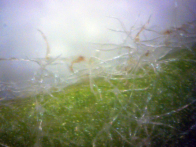
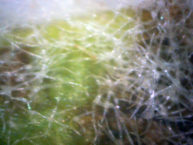
Here we see that this isn't hair at all, but hyphae, a key sign of infection.
This leaf bottom was loaded with it! (400x)
7
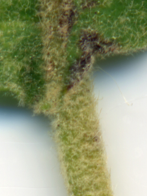
The top of the leaf also was filled with these hyphae.
But, as this picture shows, nowhere were they thicker than at the stem. (400x)
The super abundance of these hyphae leaves little doubt that this tree is in serious decline,
and why the owner may cut it down.
While the earlier pictures were taken in late May, the following set of pictures
were taken in late September.
At this time the white canker had already reproduced by shedding it's spores.
Hence, spores were hard to find.
9
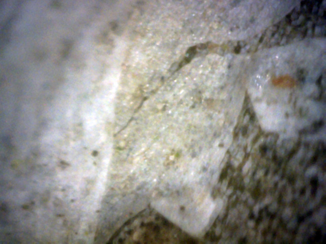
I doubt this this is white canker since this sheet-like covering isn't typical of it.
I'm guessing it's some other disease. (400x)
11
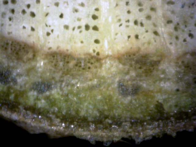
This was the best-looking twig cross-section.
Note the rich green inner bark and sapwood.
There are only a few indications of canker.(400x)
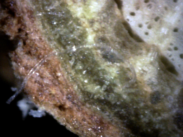
12
Lots of evidence of white canker here: it's growing on the outer bark,
a hypha ribbon is present, a white strand is in the green sapwood,
and there are voids within the sapwood.(400x)
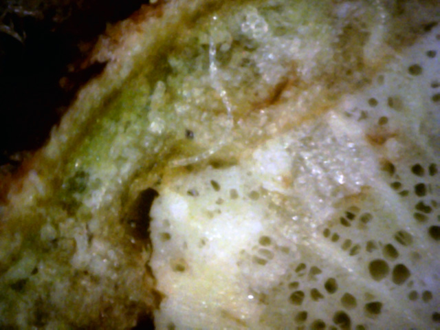
13
A large blob of white canker has invaded the green sapwood, where it has created a void.
A hypha is taking advantage of this void to spread the disease.
Even the outer bark has been mostly replaced with canker material.(400x)
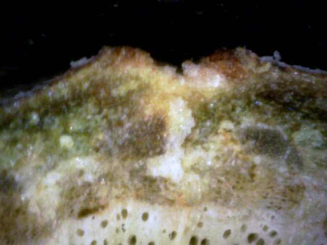
14
The blob of white canker has grown so vigorously that it has erupted through the bark.
White canker is now even growing in that wound. (400x)
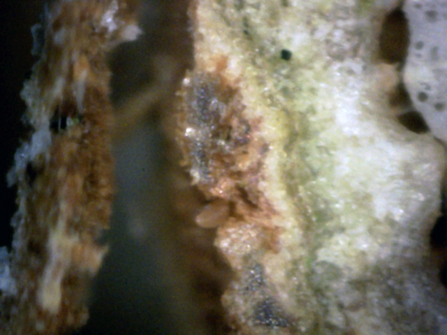
15
The line of tan white canker has so destroyed the integrity of the adjacent bark
that the outer bark has lost its adhesion.
Without nourishment, it will eventually rot and fall off. (400x)

16
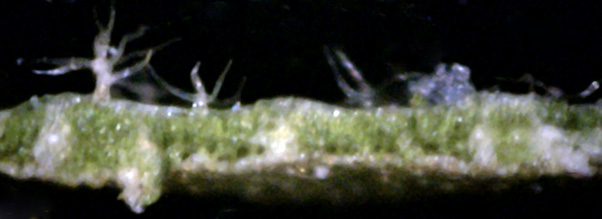
Sycamore leaves seem to have numerous spider-like objects on their surface.
This leaf cross-section shows those objects from th side.
It also looks like there is white canker material present on the bottom of the leaf. (400x)
17
Seen edge-on, those spider-like objects look like leafless, transparent trees.
There are also several white canker blobs showing. (400x)
18
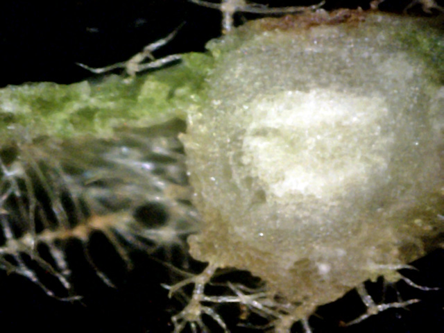
The big white blob is a leaf vein.
The tree-like hyphae seem to like being near the vein, probably because its a source of nourishment. (400x)

