With 35 years of growing experience, I've found that Mulberry trees seem resistant
to almost any disease.
Not to white canker, however.
The leaves shrivel, develop black spots and die. Then the branches die.
1
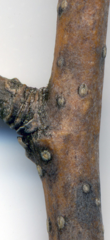
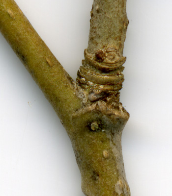
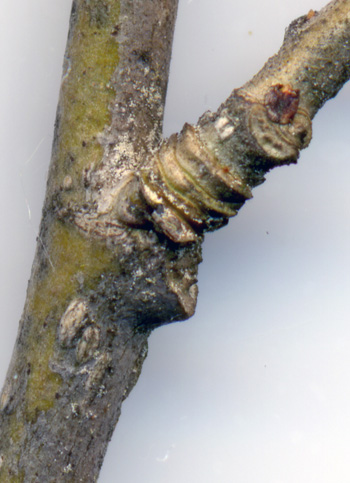
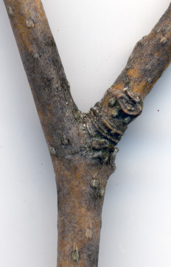
The quarter-inch branch in these three pictures no leaves.
Note the oval bark fissures in very close proximity to black bark.
Pictures 1 and 2 show spores in the node (branch junction).
Picture 3 is of a leaf scar located about 15" from the branch tip.
The area around the scar is filled with spores.
Away from the leaf scar, the spores lie mainly on top of
the black (and presumably diseased) bark.
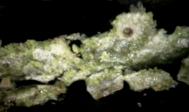
Pictures 4 and 5 are of a branch that still has leaves and fruit.
The branch shown in picture 4 is closer to the branch tip, and the one shown in
picture 5 is about 15 inches from it.
Newer growth appears more disease-free than older growth.
Picture 6 shows spores growing on the branch as well as at branch junctions.
The spore cluster locations generally seem to be from 6 to 20 inches from the branch tip,
which seems fairly typical for white canker spore production.
While the preceding pictures were taken in late May, the following set of twig
surface pictures were taken in late September using a 400x digital microscope.
 ↑
↑ 7
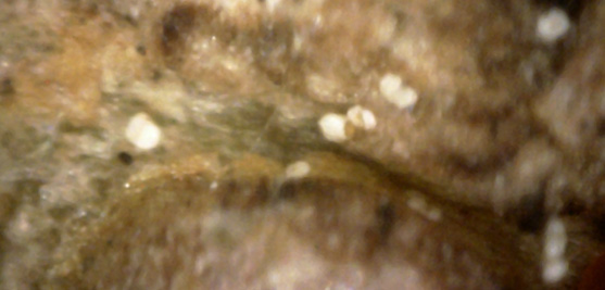
This bark surface area near the branch junction contained a fair number of spores.
Of interest here is the spore near the middle that seems to be exposing a yellow inside
(red arrow). (400x)
 ↑
↑
8
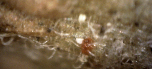
Here are two rare spherical orange objects (red arrow).
They are probably associated with the adjacent white canker objects since
they have hyphae growing through them. (400x)
9
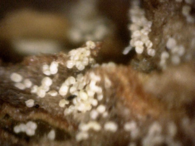
This area near the twig junction (branch bottom) had a large number of spores.
But these may be old spores.
There is a lot of semi-transparent light brown hyphae mixed in with them. (400x)
Now that we've seen what the surface looks like, the following set of pictures shows what the
twig interior looks like when making a cross-section of the twig using a 400x digital microscope.
 →
→
→
→
10
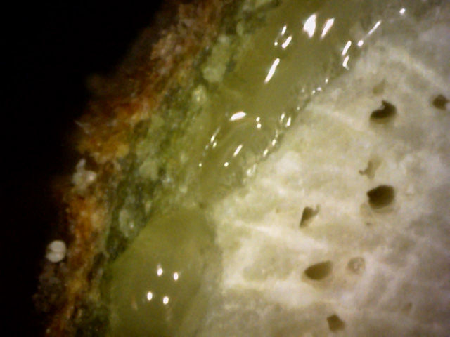
There are two items of interest here.
One is the spore in the bark cavity (red arrow).
The other is the spore below it (blue arrow), which seems to have a light yellow shroud over it,
indicating a yellow spore may be evolving into a mature white spore.
That may be true of the spore in the cavity too, although the yellow material
seems to have "spilled out" (a birth defect?).(400x)
11
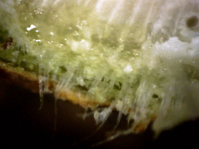
There is a massive hyphae presence in this twig tissue.
Plus, the green cortex is very fragmented.(400x)
 ←
←
12
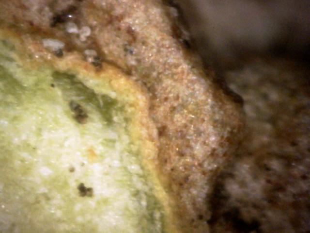
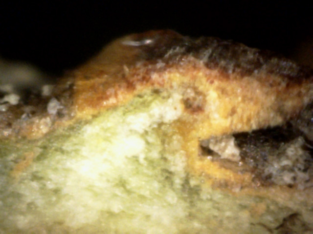
Here there was a significant white canker infection, and the tree tried to react
by building bark tissue around it, causing a growth consisting of of normal bark
and white canker. The canker did manage to eat out a void, however.
The void, along with a bark presence, apparently caused the canker to emit some
spores within the void (red arrow).(400x)
13
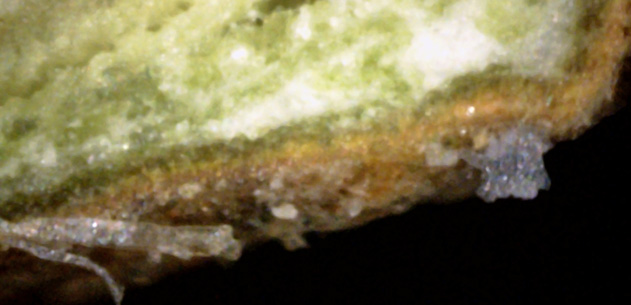
More evidence of where a white canker infection tried to push through but the tree
fought back trying to enclose it. (400x)
 ↓
↓
↑
↓
↓
↑
14
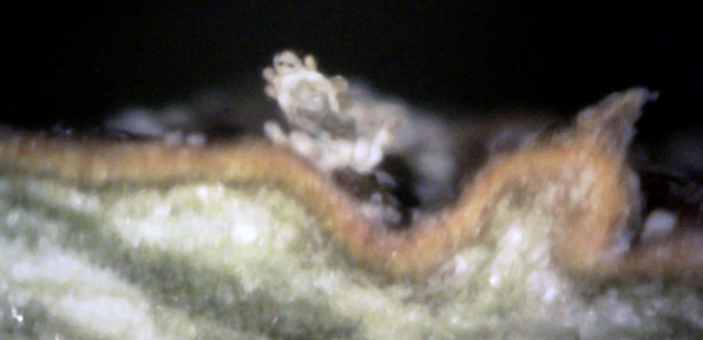
There are three components of white canker present here: a light gray blob (red
arrow), a yellow compact object (blue arrow), and a semi-transparent mass of
spherical objects (green arrow). (400x)
 →
↓
→
↓
15
When making this twig cross-section, the razor apparently cut through a blob
of white canker (red arrow), showing a snow-white shell, a gray inner hollow,
and some dark material spilling out.
But look carefully around the outside of this white shell and you can see 4 or 5
budding yellow fingers (blue arrow).
These buds may be an attempt to generate new spores, due to the presence of bark close by.
(400x)
While the wood of the tree is an excellent diagnostic tool for detecting white canker, the
leaves can also provide substantial supporting evidence. The following set of pictures
deals only with the leaves. All pictures were taken
using a 400x digital microscope.
16
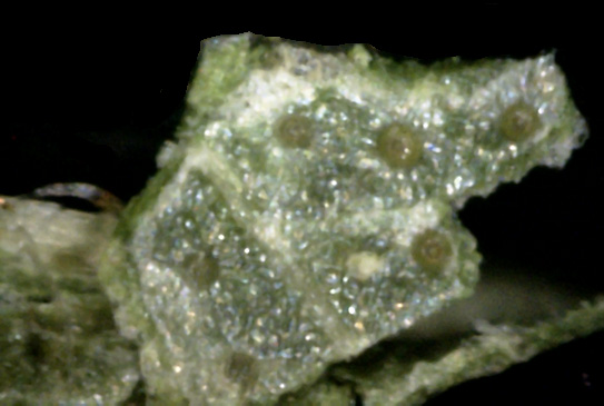
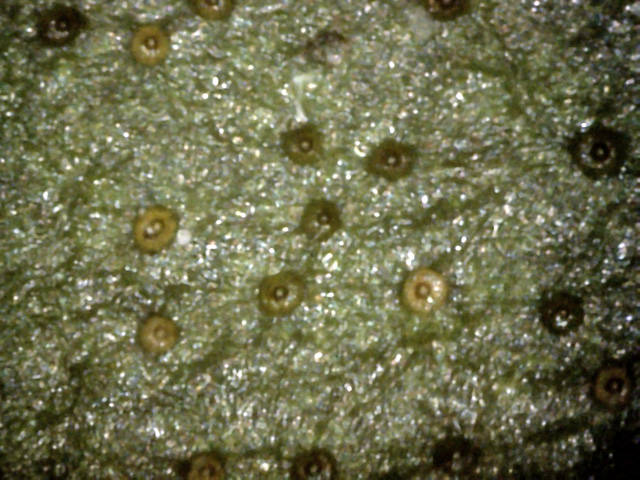
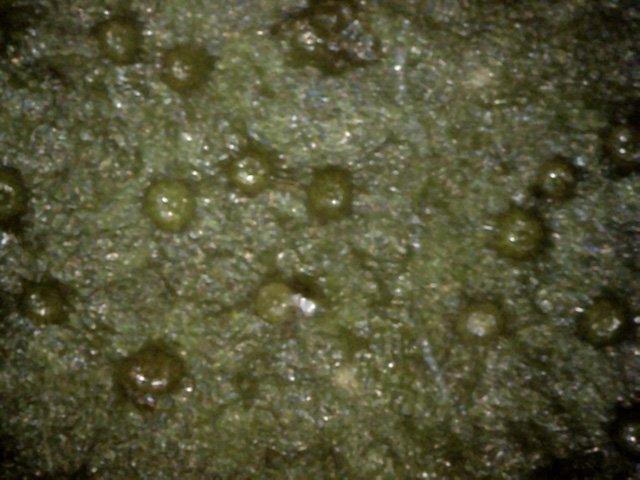
The leaf top was littered with these strange circular objects.
Curiously, the center of them was white surrounded by a ring of brown.
One had two white dots in the center.
They can't be stomata (breathing pores), since stomata are found on the bottom
of a leaf.
(400x)
 ↓
↓
17
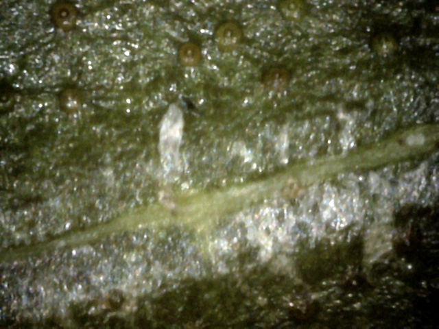
More of these mysterious round objects.
But look carefully at the one near the center (red arrow) - it seems to be sprouting a new
clear hypha near its top-center. One near the top seems to be doing the same thing.
(400x)
18
More of those olive-green circular objects.
But there is a fair amount of white canker also present, and, as I've seen before,
it seems to like to congregate near leaf veins (where the nutrients are).
(400x)
Not all the action takes place on the top of the leaf. Usually, the bottom of
the leaf is more informative about white canker symptoms than the top, as shown
in the following pictures, which were all taken
using a 400x digital microscope.
19
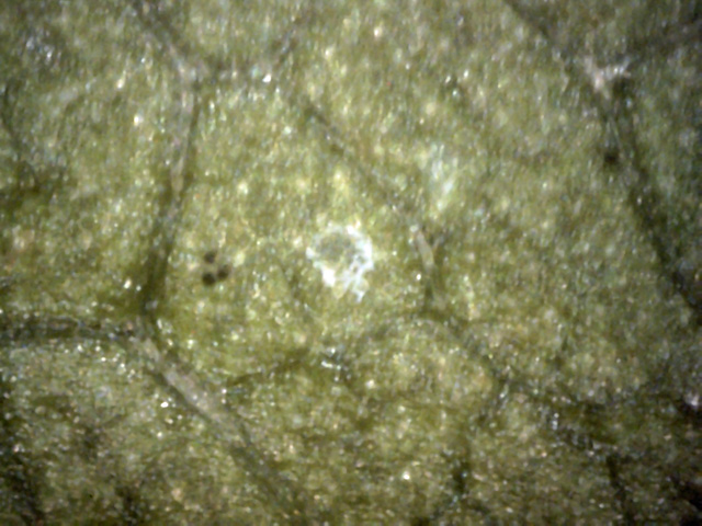
There is a diffuse blob of canker in the middle of the picture, and a more
concentrated area in the upper-right, over a leaf vein.
Most of the leaf bottom was like this - not much canker visible.
(400x)
20
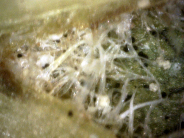
At first, I wasn't going to include this, but decided to anyway because I now
believe it says something significant.
Canker hyphae and spore generation seem to be enhanced near the surface (epidermis)
of a leaf vein or near the the surface of bark.
(400x)
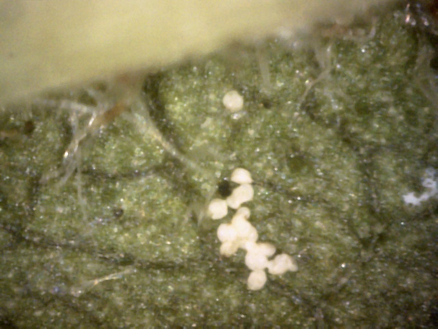
21
The leaf veins often had hyphae coming off of them.
I suspect this was canker hyphae, but I'm not sure.
The presence of these spores close to the vein and hyphae lend support
to the belief that the hyphae are canker related.
(400x)

22
The interesting thing about this rather poor image is that a "hair" coming out of
a leaf vein has a yellow blob attached to its end.
Hence, its probably not a normal leaf hair, but a hypha of white canker that
may be giving birth to a new spore.
(400x)
The analysis of the leaf wouldn't be complete unless we had a peek inside the leaf.
The following pictures show this via a cross-section. Once again, these pictures
were all taken using a 400x digital microscope.
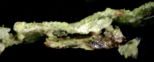
23
The big blob of canker is growing among the leaf tissue cells, almost completely
engulfing a part of it.
(400x)
