Ginkgo trees go back about 150 million years, so they've probably survived more diseases than any other tree.
This particular tree is relatively small - about 10' tall.
It was planted only a year or two ago. Yet, it is showing signs of stress.
Many of its leaves look stunted while some are almost full size.
But virtually no other leaf damage is apparent.
The branches are also surprisingly clean, and host almost no spores.
This tree was sampled in early June.
1
←
→
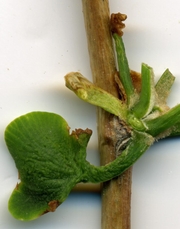
I cut off most of the leaves at this leaf node in order to be able to scan this branch, and
also to highlight the diseased leaves. The leaf on the left appears shriveled, as if badly diseased.
Two other leaves were so diseased that they didn't develop at all (black arrows).
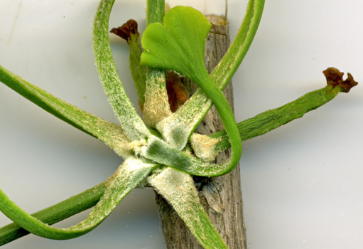
The base of the leaves at this node contain a lot of "hair".
This hair is associated with some tree diseases.
2
This is a particularly good shot of the "hair" at the leaf junction area.
Two new leaves that died during development are also shown.
3
4
→
→
→
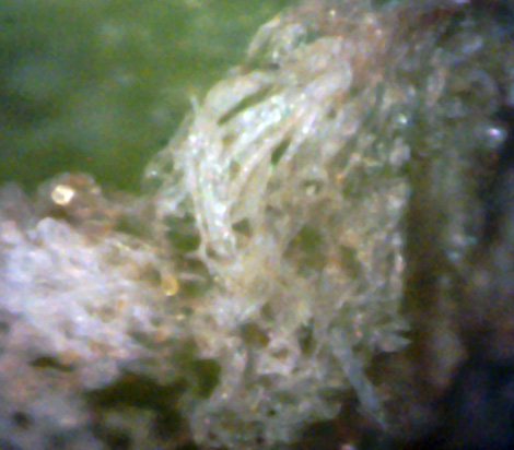
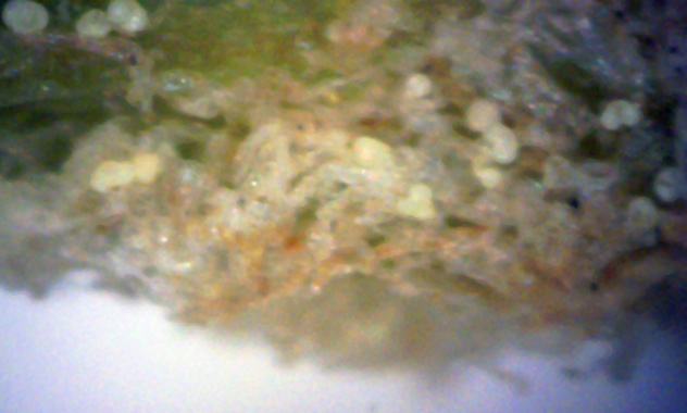
A 400x microscope view of this "hair" shows that it is really a tangled mass
of hyphae. More important, this hyphae contains both an immature spore (red arrow) and two
mature spores of white canker (black arrows).
5
6
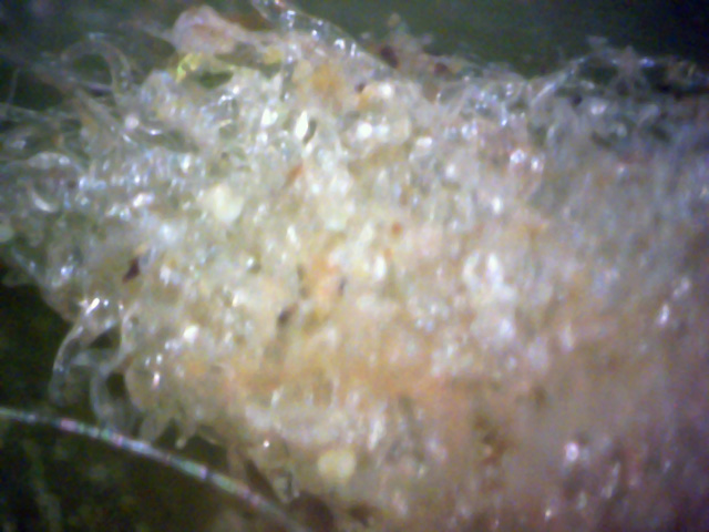
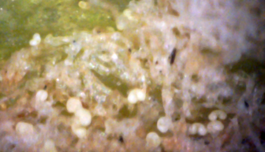
This 400x set of pictures, taken at a hyphae cluster near the leaf surface, shows
numerous white canker spores. These pictures were taken in early June, within a day or two
of a major white canker spore release. Note that these spores have a yellow tinge to them,
which is characteristic of very new spores. Some are so new, that if you look very closely,
you can even see a touch of yellow embryonic tissue between the spore lobes
(click the picture to zoom in).
Unlike other trees, the Ginkgo seems to be trying to prevent white canker spores from emerging
through its bark and propagating. In turn, white canker spores have found that they can grow out of
the leaf junction area, where there is no bark. As a last line of defense, the Ginkgo seems to be
throwing up a tangle of hyphae to trap the emerging spores so that they have trouble propagating.
The destructive part of the white canker infection is lurking under the bark, within the tree tissue,
slowly destroying it.
I have more than an average interest in Ginkgo trees, partly because their leaf shape is so
distinctive and partly because they have been around so long its like looking at a living fossil.
Although ginkgo trees are attractive (not the female trees, since their fruit is foul smelling),
they are not widely planted. But I found a group of about 7 young trees in Evergreen cemetery
up in Portland Maine. I've been following their health for several years, and noted a progressive
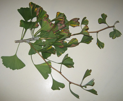
decline, with the symptom being leaf spots. So early this October I clipped a twig whose leaves showed
such spots and brought it home for analysis. The results are shown below.
1
Of all the diseased branches on this group of ginkgo trees,
this branch seemed to be the most diseased. But they all shared the same leaf
spot infection symptom. Interestingly, the vast majority of trees in this
cemetery are maples, and maple trees seem very susceptible to getting and
spreading white canker.
2
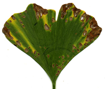
A sample leaf showing lots of disease symptoms. Spores can be seen in the dead areas.
This is the leaf used in the leaf surface and cross-section views below. (The circular
arc impressions were from my examinations with a digital microscope.)
(1200 dpi)
The next set of pictures shows the top of the leaf. The numerous dead and discolored
areas hint at severe leaf damage, and the following microscopic pictures detail
exactly what this damage is.
 ↓
↓
3
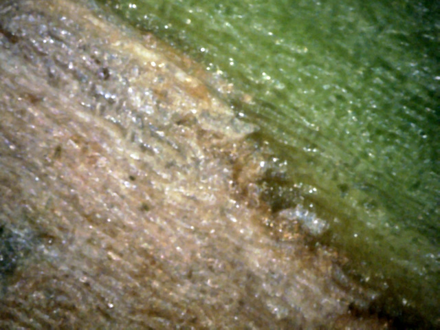
Notice the dead white area and the white canker at the edge.
One hypha is poking through a hole (red arrow).
Most of the normal green leaf looked like the surface in the upper right corner.
(400x)
 ←
↑
←
↑
4
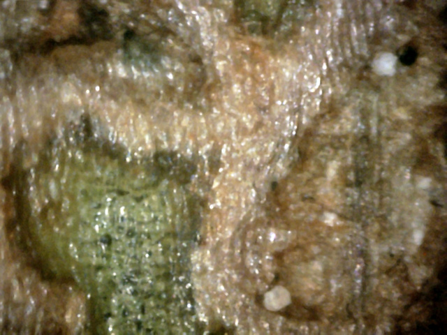
A clean area is surrounded by a branching white canker infection.
There are two white spores (red arrows) adjacent to this "river of canker".
(400x)
5
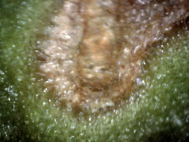
This picture reminds one of an explosion in a liquid, with the hyphae of white canker
representing liquid streams splashing from the center finger-shaped dead leaf area.
(400x)
The leaf bottom can also give some insight as to what is going on. The next two
pictures show the leaf bottom. In particular, the contrast between the live and
dead areas is of interest.
6
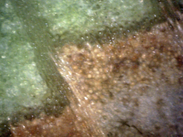
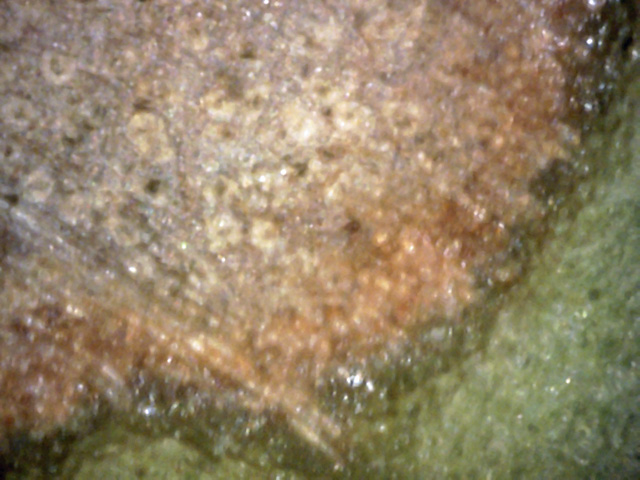
The edge of a dead area.
The white blobs seem to be either particles of white canker or
leaf cells that have been overrun with white canker.
Notice that the white canker infects the leaf vein as well as the leaf cells.
In fact, you can see the canker tentacles spreading out from each tubule
in the leaf vein if you look closely.
(400x)
7
An advancing front of dead leaf tissue.
The ghostly white blobs in this dead area may be leaf breathing holes (stomata)
that were infected and killed.
(400x)
Somewhat surprisingly, leaf cross-sections seem to provide some of the most
informative data regarding white canker infection. Specifically, it is the area
between relatively healthy leaf tissue and diseased dead tissue that shows us
detailed white canker information, as shown below.
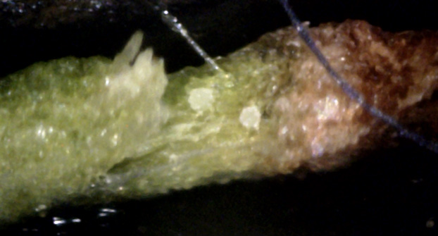
8
An infected and torn leaf vein appears on the left.
To its right are two canker particles on small stalks,
and immediately to their right is a area being killed by white canker.
Killed leaf material is shown to the right of that.
(400x)
 ↑
↑
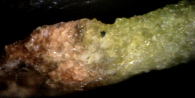
9
Relatively healthy green leaf tissue on the right.
Dead, shrunken leaf tissue on the far left.
The battle line is in between, which is swollen with white canker material.
Notice where the razor cut off a white canker white hypha (red arrow).
(400x)
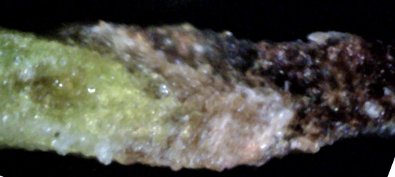
10
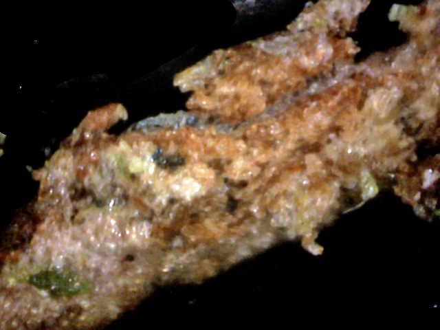
Another leaf infection boundary where the white canker growth is consuming
the healthy leaf tissue.
The leaf cells seem to turn transparent in the initial infection stage, then tan,
and then black.
(400x)
 ↑
↑
11
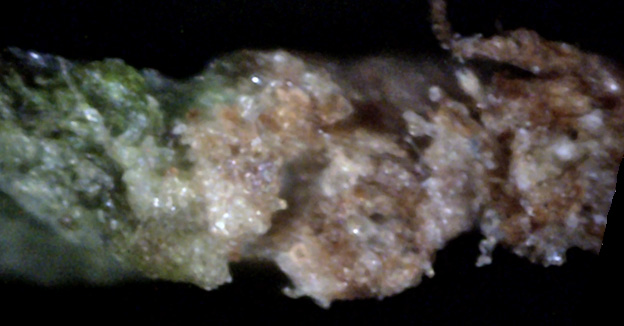
A good title for this picture might be "white canker gone wild", since it seems to
have completely destroyed this part of the leaf with various forms of canker material.
Note also the hypha snaking through the dead leaf tissue (red arrow).
(400x)
 →
→
12
Another white canker infection front.
A big hypha can be seen on the edge of the green tissue on the left (red arrow).
Numerous smaller white canker growths can be seen on the right.
The most vigorous white canker growth is seen where the green leaf tissue ends.
After the white canker has killed the leaf tissue, it seems to start sending
out transparent hyphae to find other tissue to infect.
(400x)
The most consistantly reliable way of detecting white canker appears to be the
microscopic examination of a twig cross-section. This can show the presence of
white canker even if there is no evidence of it when looking at the bark or
leaves. So while the preceeding pictures give strong confidence of the presence
of white canker, the following twig cross-section pictures pile on even more
evidence of its presence.
 ↑
↑
↑
↑
13
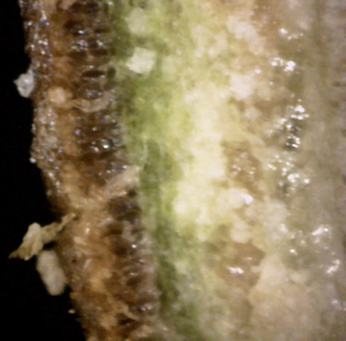
Lots of canker here.
In fact, you can see one plume of it rising up and through the bark and giving
rise to a spore (which appears to be ruptured) (red arrows).
Another plume is trying to break through the bark.
And the brilliant white area indicates that there is lots of canker growth in the phloem,
the energy-rich part of the wood.
(400x)
14
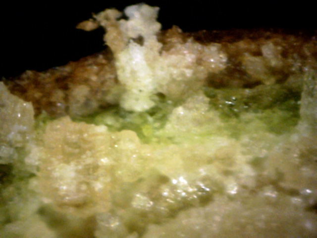
There are huge billows of canker growth in the phloem.
Also, a large body of canker is pushing its way up through the bark.
The bark appears to be about 80% destroyed in this area.
(400x)
15
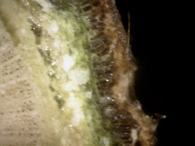
The beige colored area on the far left is the xylem, and, like other trees,
it is virtually unaffected by canker growth.
But to the right is the phloem, and the brilliant white blobs show that white canker
is rampant throughout it, so much so that the phloem is separating from itself
and from the xylem.
Except for the extreme outer surface, the outer bark has been almost totally
infected with canker.
(400x)
16
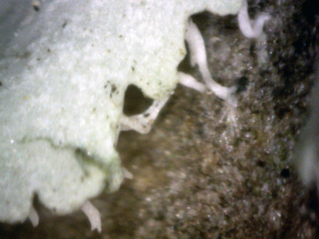
This is what lichen looks like.
Notice that the appearance is different from canker.
Lichen has a blue-green tinge to it, while white canker is white or light gray.
(400x)
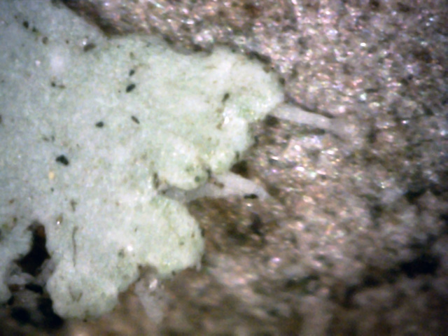
17
Lichen roots can be seen digging into the bark.
The lichen surface also seems to have a lot more debris on it than a canker would.
(400x)
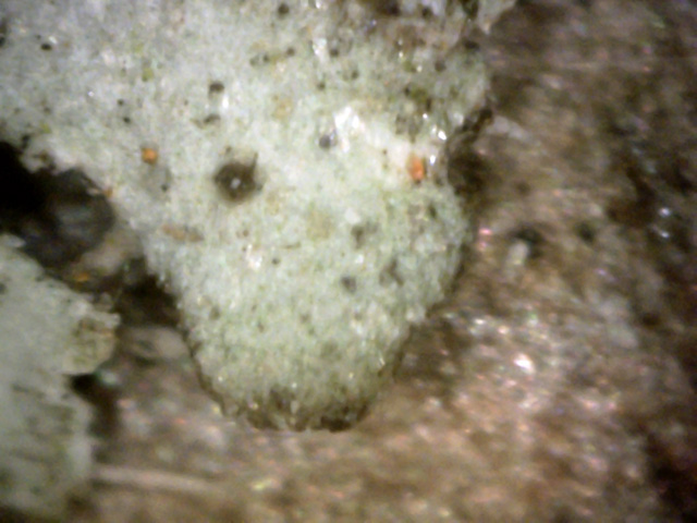
18
A tip of blue-green lichen on a quarter-inch thick branch.
(400x)
Recall from the first picture that there was some lichen on the branch. For
comparison purposes, here are some pictures of that blue-green lichen using a
digital microscope set to 400x magnification.


