Our yard contains both a small "Stellar Pink" Rutger’s hybrid dogwood tree
and a larger Kousa hybrid dogwood tree. Both are hybrids that are claimed to be resistant
to anthracnose, which is killing off native dogwoods. But both of these trees have shown
disease symptoms during the past few years (starting about 2004).

1
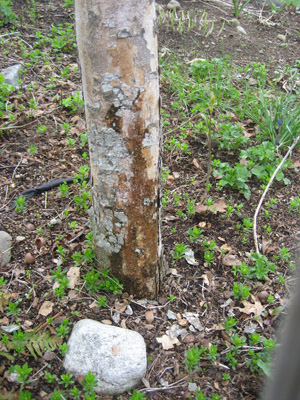
This is the trunk of the Stellar Pink dogwood that underwent a microscopic exam in the following pictures.
That long vertical split developed soon after I bought the tree several years before this picture
was taken. While it appears to be due to mechanical damage, the split was instead caused by White Canker
disease. A fungicide treatment resulted in healing. But no fungicide treatment had been applied
here for the past month, and as you can see, patches of bark are falling off. The bark is also supporting
some kind of light gray fungal growth. This tree is in a severe struggle to survive.
2
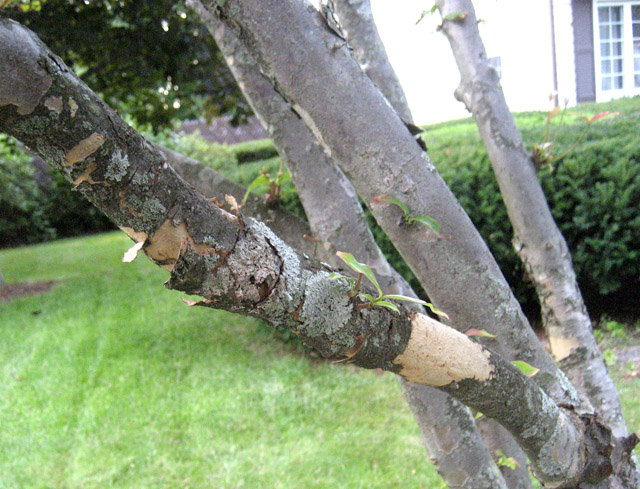
This is another Dogwood tree in our yard - a Kousa variety, that is a disease resistant hybrid.
In fact, it did fine until a few years ago when White Canker hit it.
Notice that almost all of the outer bark on the trunk has fallen off, and sap is seeping through the inner bark - not a good sign.
Only a fungicide treatment has saved this tree.
3
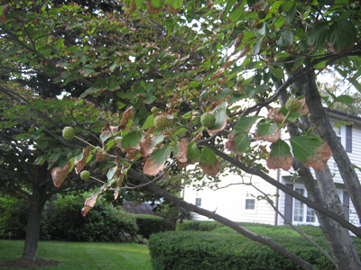
This Dogwood tree belongs to a neighbor about a block away.
The severe bark shedding shows that it, too, is in serious decline.
4
This is the foliage on that Dogwood with the severely peeling bark shown
in picture 3.
The leaves appear to be having trouble getting enough moisture.
6
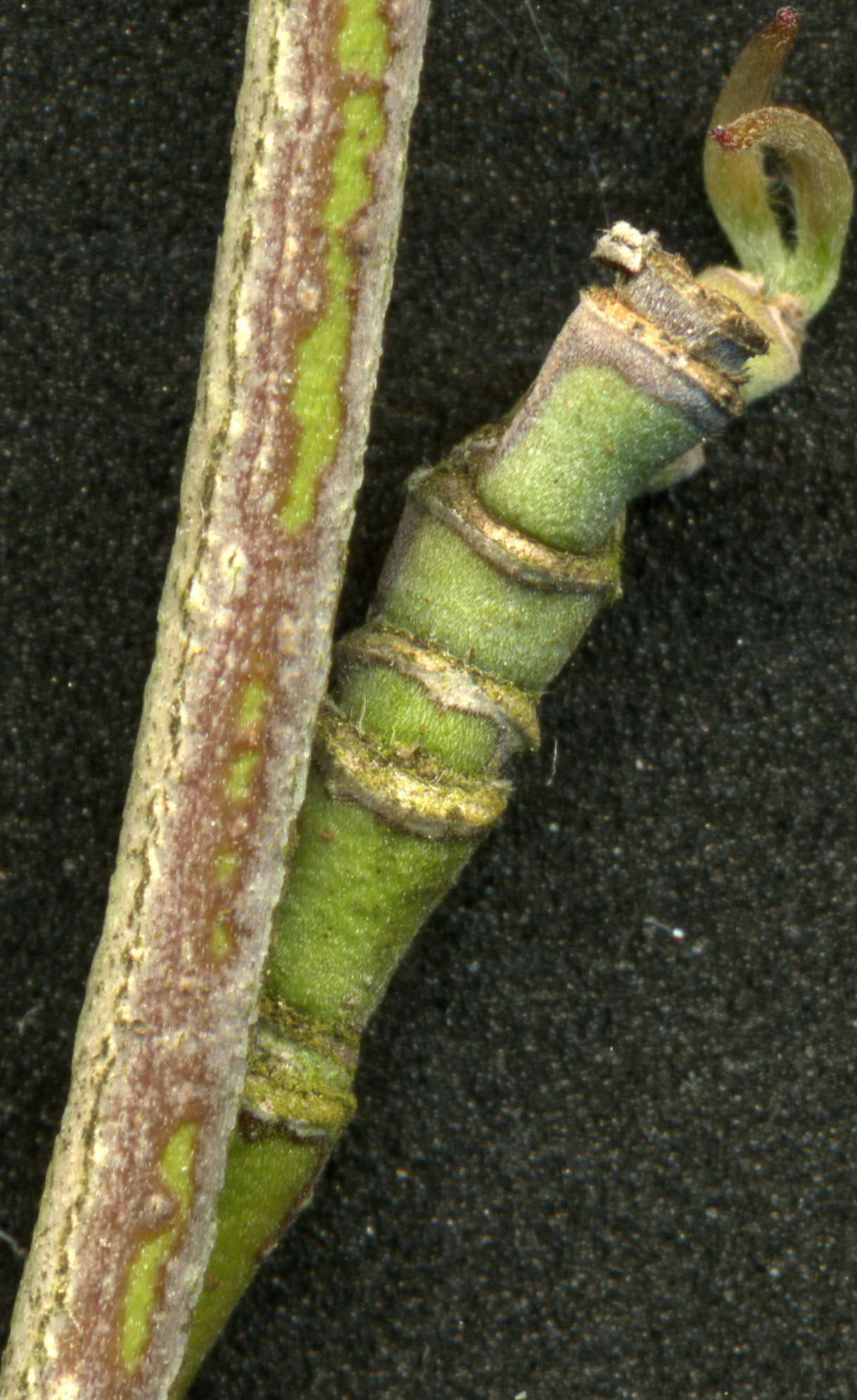
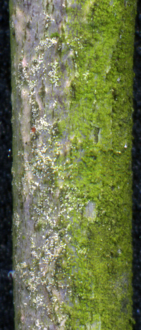
Pictures 5 and 6, along with the following pictures, were from the stellar pink
dogwood shown in picture 1. Pictures 5 and 6 are of the same branch, scanned at 2400 dpi.
While picture 5 shows the tip of a branch, picture 6 shows the bark further on down toward the trunk.
The terminal growth shown in picture 5 is distorted, indicating an infection.
Picture 6 shows that the slightly older growth contains an abundance of yellow spores.
Extensive lichen growth is also present near the spores.
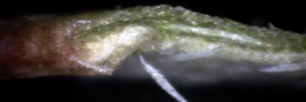
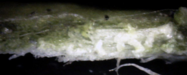
Suspecting the leaves might show some disease evidence, I placed a diseased leaf from this tree in a vise
and used a razor to slice off the protruding piece.
A 400x digital microscope was then used to photograph several leaf cross-sections, as shown below.
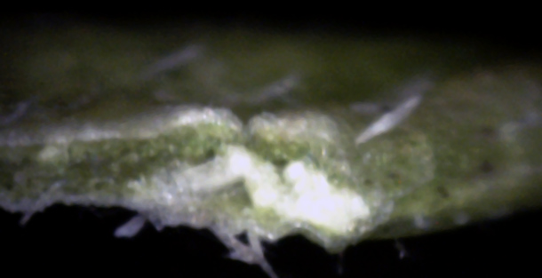
11
Another patch of white canker growth accompanied with hyphae growing through the lower leaf surface.
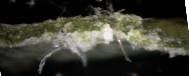
12
More white canker on the lower leaf surface, along with its hyphae.
Note that one patch of hyphae is also growing out of the top of the leaf, and may also extend
out of the bottom of the leaf.
In general, most of the white canker is buried within the leaf, and tends to be milk-white in color.
This color fades to a translucent light white (or gray) for canker material on or near the surface.
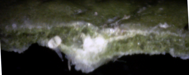
As informative as these leaf cross-sections are, twig cross-sections are often even more useful, as shown by the following 400x microscope pictures.
As informative as these leaf cross-sections are, twig cross-sections are often even more useful, as shown by the following 400x microscope pictures.
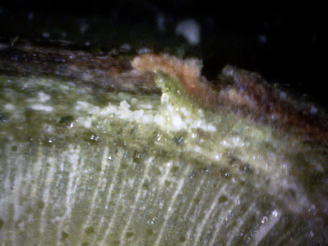
13
The phloem here has a relatively large area of embedded white canker growing up through it.
Notice that where the white canker density is highest, the bark next to it has lifted and
turned a "peanut butter brown" color.
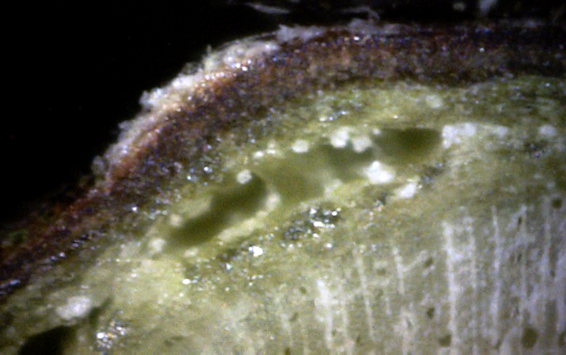
14
↑
↑
The growth of the white canker under the bark has split the phloem layer, creating voids (red arrows), and
cutting off the bark from nourishment. The bark here is bulging out, and white canker material
is growing on that bulge. The dark brown bark will mix with the white of the white canker, giving
an external visual appearance of light brown.
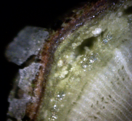
15
Here again, the white canker growing under the bark is creating a void by splitting the
phloem layer and causing the bark to bulge out.
Notice that the canker material near the void is tending to form a spore-like object.
Also notice the large blobs of white canker material growing on the surface of the bark.
In summary, the distorted new growth and the large number of tan spots on a new branch give strong
hints of white canker. The leaf cross-sections reinforce this diagnosis.
The strongest evidence comes from a twig cross-section, where the white canker can be seen growing
in the nutrient-rich phloem, splitting the phloem, pushing the bark out, and causing white canker to
grow on the bark's surface. Finally, internal white canker tends to be milk-white in color and opaque,
while external white canker tends to be a translucent light-gray.
A very interesting footnote: A leaf from this obviously diseased tree was sent to a state plant diagnostic lab, but
no pathogen diagnosis was found! However, a Bartlett Tree Research Laboratories website
document stated:
"Approximately seventy percent of the dogwood canker and root samples received ... are confirmed positive for Phytophthora infection."
They later added that "Phytophthora trunk cankers are typically located near the ground.
Bark falls away after death of the infected area and may show ridges of callus tissue.
Dark liquid may be oozing from the margins of the canker where the fungus is still active.
The wood beneath the active area is dark reddish brown and watersoaked."
That description seems to exactly fit with what I'm seeing. It also goes along with my guess that
White Canker is a Phytophthora disease. Unfortunately, Bartlett Tree did not identify the species of Phytophthora
although some other sources have said this is "Crown Canker" caused by Phytophthora Cactorum.
