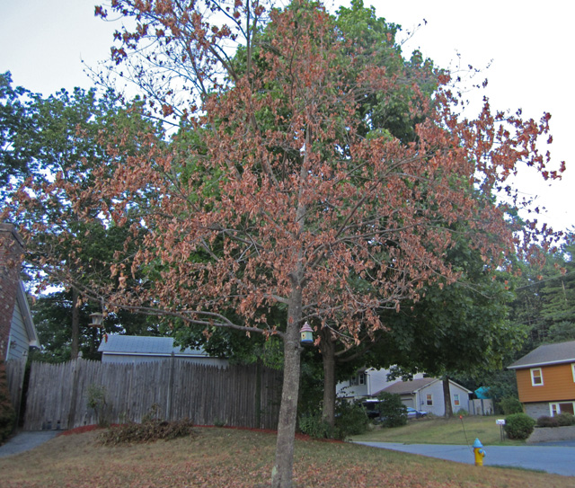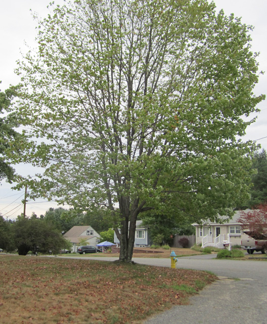
Although there is only a dead branch or two, as you can see, the foliage is very sparse, and many leaves have already fallen. Curiously, the right side of this tree is mainly south-facing, so gets most of the sun. Apparently the sun's energy is helping keep this tree going.
Except for the leaves drooping and being rather small, I saw no canker on the top or bottom surface of them.
But the bark was another matter. Actually, as is typical, most of the bark is clean. However, there were extensive patches of spores at the underside of branch junctions, as shown in the following branch surface microscope photo taken at 400x:
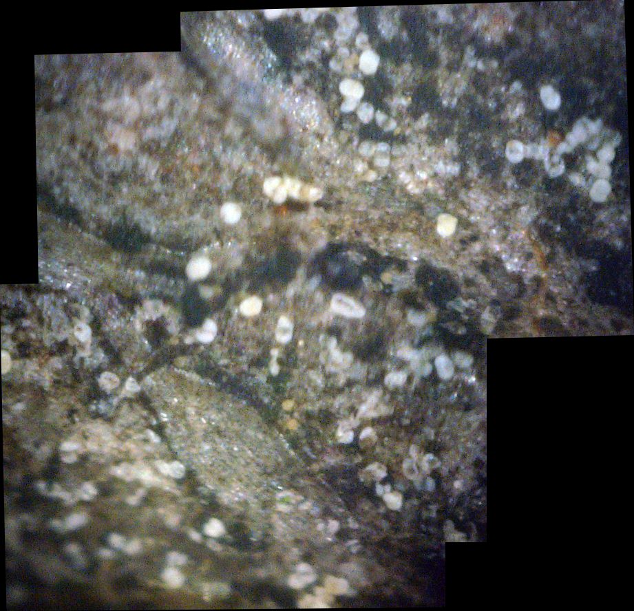
Once again, the white spores are about 50 microns in diameter and the smaller yellow spheres are about half that size.
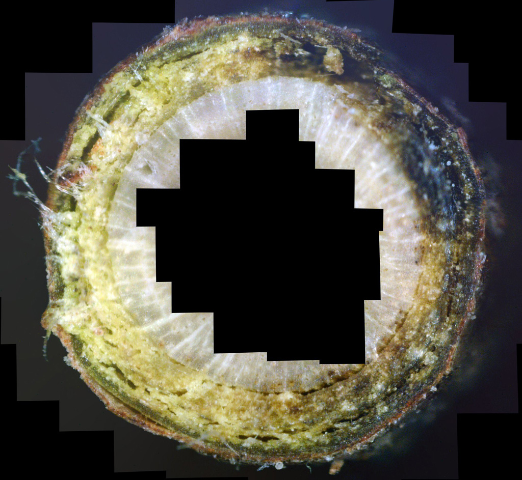
If white canker is really present, some of the best evidence is a detailed view of the cross-section of a twig or small branch. This time I took a quarter-inch diameter branch as a sample. Using my digital microscope, I then carefully took about 60 overlapping photos at 400x around the edge of the cross-section so I could capture all of the area just under the bark. I then used Microsoft's ICE photo stitching program to compose one large image. A small view of it follows:
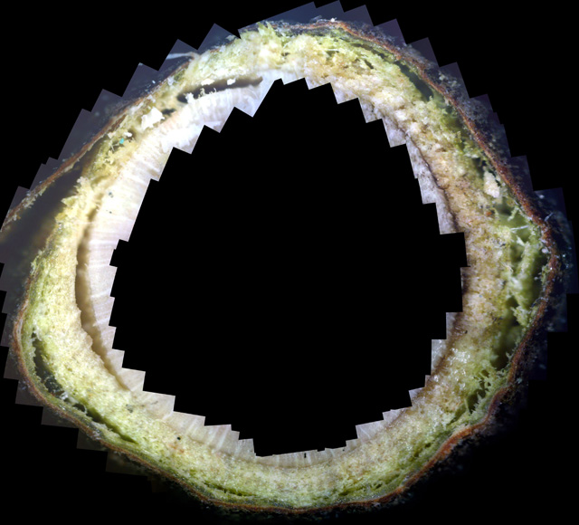
This photo shows of how badly this tree is diseased. Only a trace of the normally green phloem is present, having been replaced by white canker material. The bark is separating from the phloem, as if it is trying to get away from it. Only a very thin layer of phloem remains immediately under the bark, and even there it only extends about half-way around. I've also made the full-resolution photo available on this website - check it out by zooming into it, at: www.whitecanker.net/pictures/2010-07-24 Maple branch cross-section donut.jpg
(Right click the picture to save it to your PC or to print it.) As you zoom-in to examine the details, you'll gain a better appreciation of how devastating this disease is to the inner part of the tree. The chaos there reminds one of a war going on. And, in fact, there is.
You'll note that these photos don't include the central xylem, which is usually a boring white. You can see the edge of it in the above photos. However, white canker does infect this xylem. Apparently it has killed part of the xylem in the center of this branch, as the following stitched photomicrograph shows:
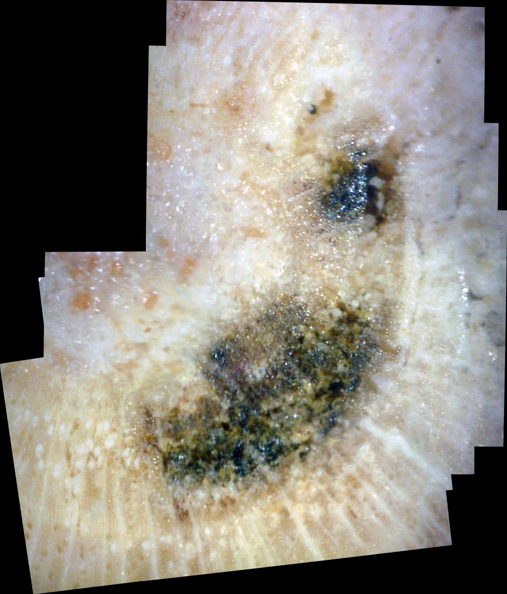
The reason I suspect white canker killed this tissue is that there appears to be white canker within and around this black tissue. Not only that, but to the lower-left of this dead area is a patch of white canker blobs. Apparently, when the white canker density gets high enough, it kills off the white xylem tissue. Furthermore, as the snow-white xylem begins to die, it seems to turn brown, further highlighting the infiltration of white canker blobs.


 I began my treatment study by having a commercial lawn & tree care company do my trees. Of the four fungicides they tried, only Propiconazole and Mancozab were effective.
